Cutting-Edge Eye Care Tech at Drs. Ditto and Musick Eye Care Center, Nicholasville
At Drs. Ditto and Musick Eye Care Center, we provide advanced and effective eye care. Our practice has cutting-edge technology that assists us in diagnosing, treating, and managing a wide range of eye diseases and visual conditions.

Innovative Instruments for Precise Diagnosis and a Customized Approach
The high-tech instruments in our practice boost our commitment to your eye health. They allow us to offer accurate diagnoses and tailored treatment plans, ensuring you receive the highest level of care.
Please note that we do not use all instruments for each patient. Every individual's needs are unique, so we will specifically tailor the instruments chosen for your exam to your condition.
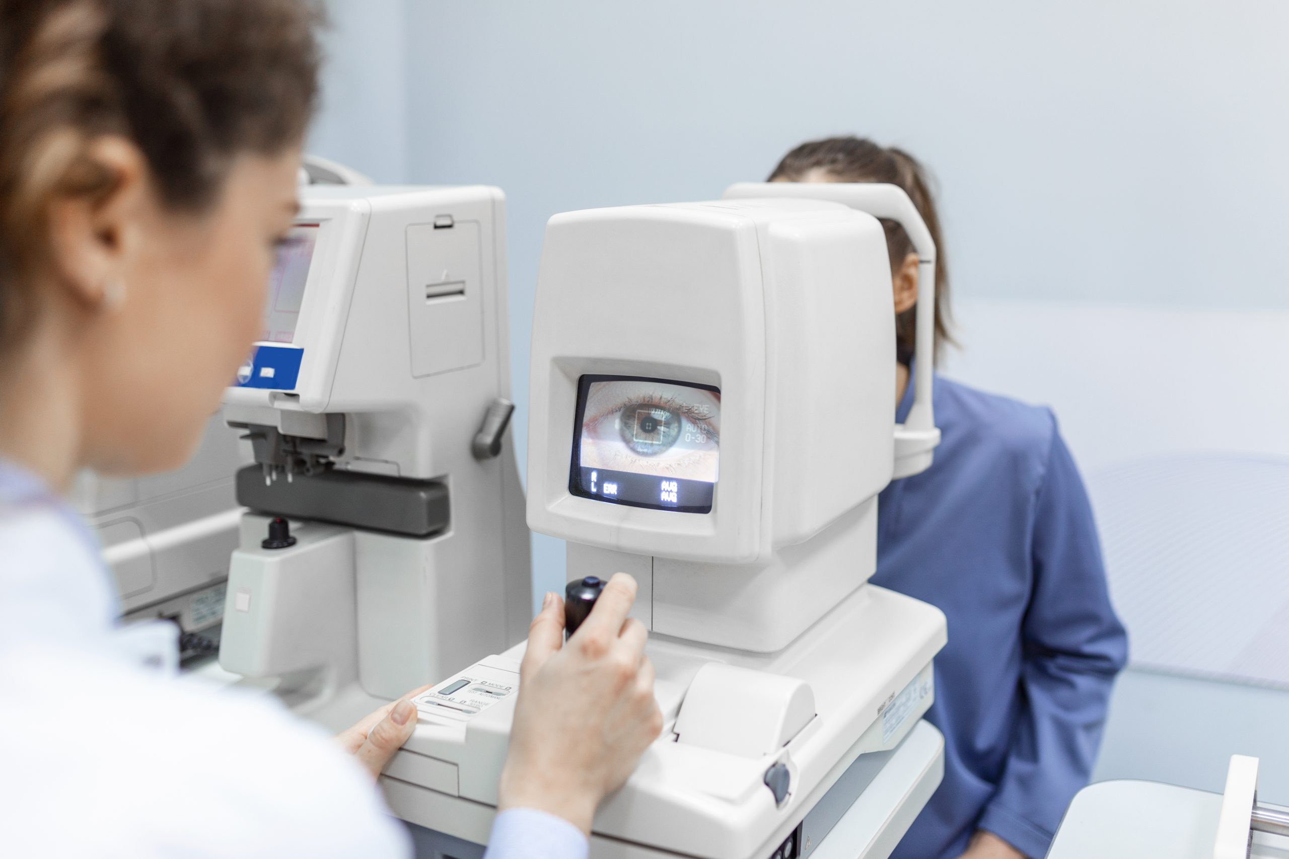
NIDEK OPD-Scan III Wavefront Analyzer
The NIDEK OPD-Scan III is a revolutionary vision-assessment system that combines corneal topography, wavefront analysis, autorefraction, keratometry, and pupillometry. This comprehensive approach allows for accurate evaluation of corneal and optical aberrations, providing a clear insight into your eye's health.
Key Features:
- Point-by-point eye aberration mapping with OPD Map
- Precise mapping of irregular astigmatism due to increased data points
- Unveiling pathways for vision correction
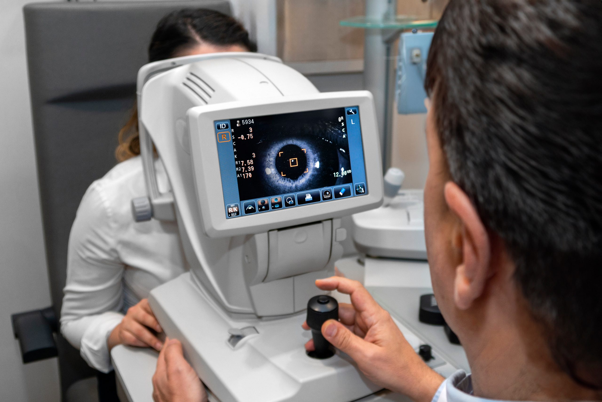
REICHERT Ocular Response Analyzer® G3
The REICHERT Ocular Response Analyzer® G3 is a tonometer that measures Corneal Hysteresis (CH), an indicator of corneal biomechanical properties. This measurement is a superior predictor of glaucoma progression, offering valuable insights for optimal management and care.
Key Features:
- Measures Corneal Hysteresis, indicating glaucoma progression risk
- Provides Corneal Compensated Intraocular Pressure (IOPcc), a more accurate pressure measurement
- Enhances glaucoma management with biomechanical insights
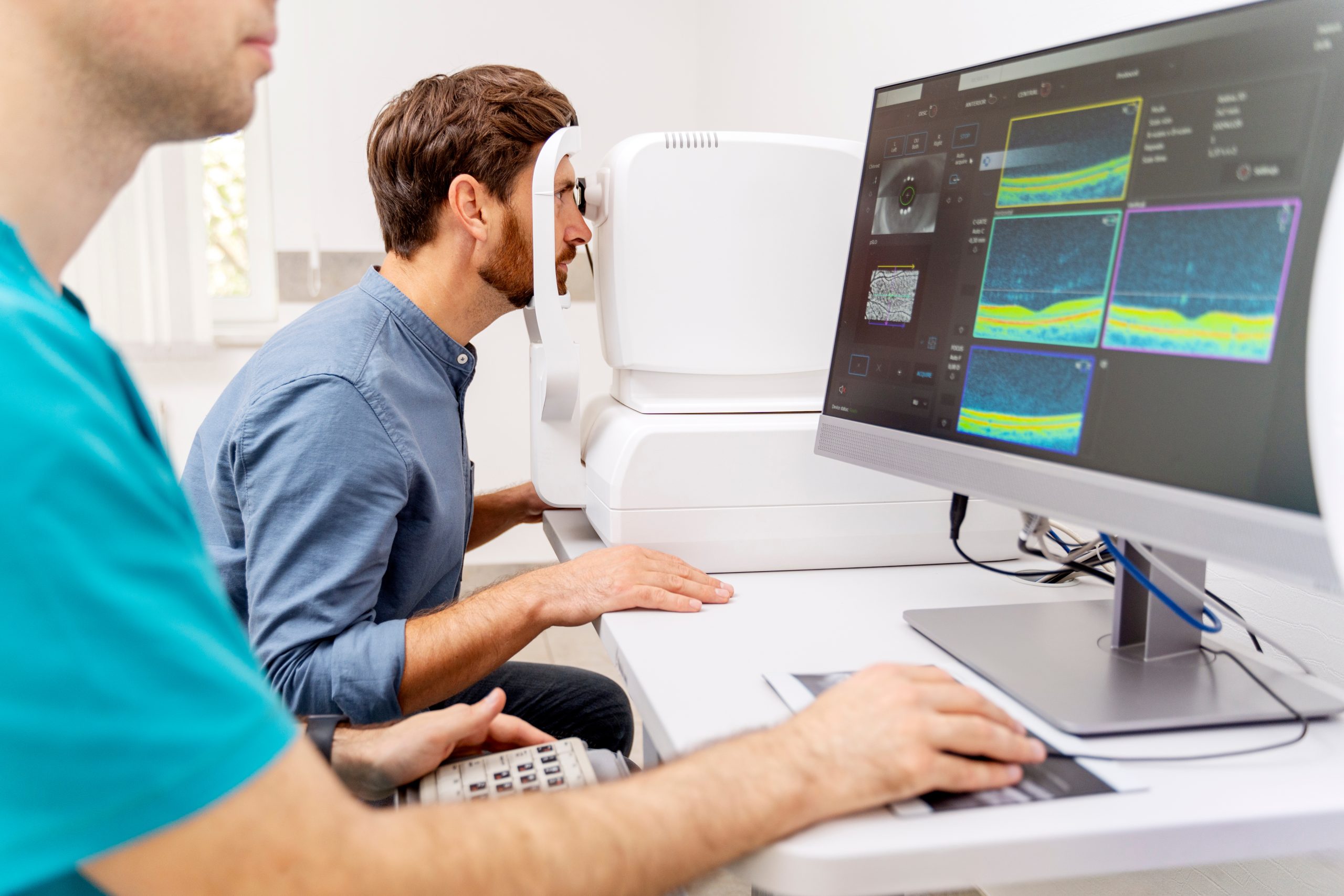
Zeiss Cirrus HD OCT
The Zeiss Cirrus HD OCT is an advanced spectrum domain OCT instrument that provides high-quality imaging for diagnosing and managing a wide range of glaucoma and retina conditions. Its precision imaging aids in accurate diagnosis and effective treatment planning.
Key Features:
- High-definition imaging for accurate diagnosis
- Valuable insights into glaucoma and retina conditions
- Spectrum domain OCT technology for enhanced accuracy
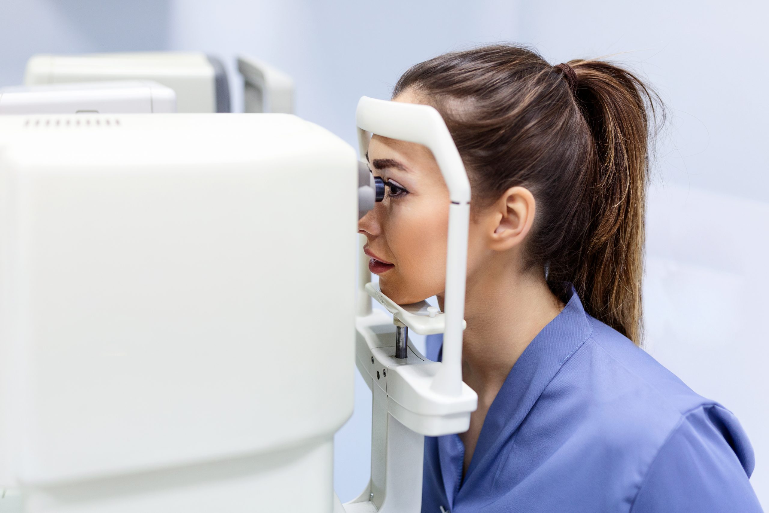
ZEISS Visucam High-Resolution Retinal Camera
The ZEISS Visucam High-Resolution Retinal Camera produces detailed images to aid in diagnosing and monitoring various eye diseases. This camera offers enhanced diagnostic insight for comprehensive eye care, from glaucoma to macular degeneration.
Key Features:
- High-resolution fundus imaging for detailed analysis
- Versatile imaging modes for diverse diagnostic needs
- Seamless integration into clinic workflow for enhanced practice performance

LIGHTMED Deux SLT/YAG Combination Laser
The LIGHTMED Deux SLT/YAG Combination Laser offers a range of in-office laser procedures, from YAG capsulotomy for secondary cataract formation to Selective Laser Trabeculoplasty (SLT) for primary open-angle glaucoma. This advanced laser technology provides cost-effective solutions with minimal disruption.
Key Features:
- Superior Crystal-Q Switch Laser Cavity for precision
- Convenient slit lamp integrated system
- Diverse laser procedures for various eye conditions